Cell Marker
The Introduction of Cell Marker

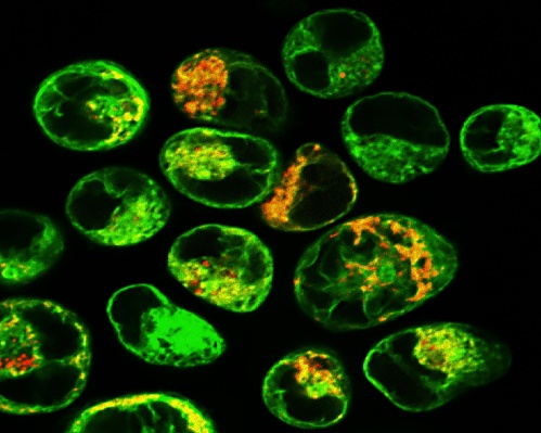
Cell markers refer to a type of marker that is specifically expressed at a specific time in a specific cell, reflecting the growth and differentiation of cells, and can be used to identify specific cells and monitor cell growth and differentiation. Cell markers are now also used in the diagnosis and treatment of many diseases.
The Research Status of Cell Markers
Cell markers have been widely used in laboratories and clinics and play an important role in the research. As one of the common markers of ovarian cancer stem cells, CD133 is a membrane glycoprotein encoded by cd133 / prom-1 gene, which has been proved to be a marker molecule of neural stem cells and hematopoietic stem cells. CD133+ cells can promote the production of vascular system and participate in the generation of new blood vessels, and play a promoting role in the growth, proliferation and infiltration of ovarian cancer cells. Baba T et al. first detected CD133+ cells in ascites of ovarian cancer patients in 2009 and confirmed that CD133+ cells had greater tumorigenic ability and tolerance to chemotherapy than CD133- cells. It was found that CD133 was highly expressed in oocyte carcinoma tissues, and CD133+ cells were more prone to tumorigenesis and migration than CD133- cells and could reproduce the original heterogeneous tumors. CD133+ was more resistant to chemotherapy drugs, and the expression of CD133 was significantly increased in patients with recurrent platinum resistance. MSCs (Bone marrow mesenchymal stem cells) are a hot topic of research in recent years. It differentiates into bone marrow cells, fat cells and chondrocytes in the body, which brings hope for the treatment of various refractory diseases. As surface markers of MSCs, CD molecules have been widely used in basic research. Currently, positive surface markers recognized by most scholars are CD44, CD90, CD29 and CD105. In addition, under the influence of tumor environment, MSCs surface markers are not absolute. For example, CD166 is expressed in normal gastric mucosa, adenoma and adenocarcinoma tissues, and CD271 is almost not expressed in normal gastric mucosa, adenoma and adenocarcinoma tissues, so it cannot be used as a specific marker of gastric cancer tumor cells. In the study of co-culture of MSCs with microenvironment of colon cancer, it was found that MSCs can be induced to transform into colon cancer cells. The expression rates of CD13 and CD133 on the surface markers of MSCs were 89.7% and 90.5% respectively. Recently, some progress has been made in the field of markers of neural stem cells. Musashi1 is an evolutionarily conservative RNA-binding protein, which plays an important role in maintaining the status of stem cells. Musashi1 is selectively expressed on nerve precursor cells, and besides the nervous system, Musashi1 is also a selective marker of intestinal stem cells. The expression of Musashi1 in stem cells or immature cells of these tissues indicates that Musashi1 plays an important role in maintaining the undifferentiated state of these cells in the post-transcriptional gene regulation stage. One target molecule of Musashi1 in the body is M-numb mRNA, which plays an important role in neural differentiation. Studies show that mutation Musashi1 inhibits the synthesis of M-numb. Numb is an evolutionally conservative intracellular Notch antagonist. So, it is speculated that Musashi1 is a positive regulator of Notch signaling pathway. Although cell markers have been widely used in the laboratories and clinics, further research is needed to find a single, specific cell marker.
References:
- Redl H, Schlag G. Lung-converting-enzyme as a possible cell-maker in connection with the ultrastructural morphology in hypovolemic-traumatic shock. Der Anesthetist. 1980, 29(10):552.
- Shimomura O, Oda T, Tateno H, et al. Abstract 5231: Undifferentiated cell maker rBC2LC-N lectin have high affinity to pancreatic cancer cells and residual cancer cells. Cancer Research. 2017, 77(13 Supplement):5231-5231.
- Anonymous. Fuel-Cell Maker Ballard Cuts Expenses, Losses. Transport Topics. 2003(June).